
Panoramic and Periapical X-rays: How Is Care Planned?
Previous images, symptoms and planned treatment are considered. Relevant health circumstances should be discussed before imaging.
Imaging is selected to answer a clinical question, and previous images may help avoid unnecessary repeats.
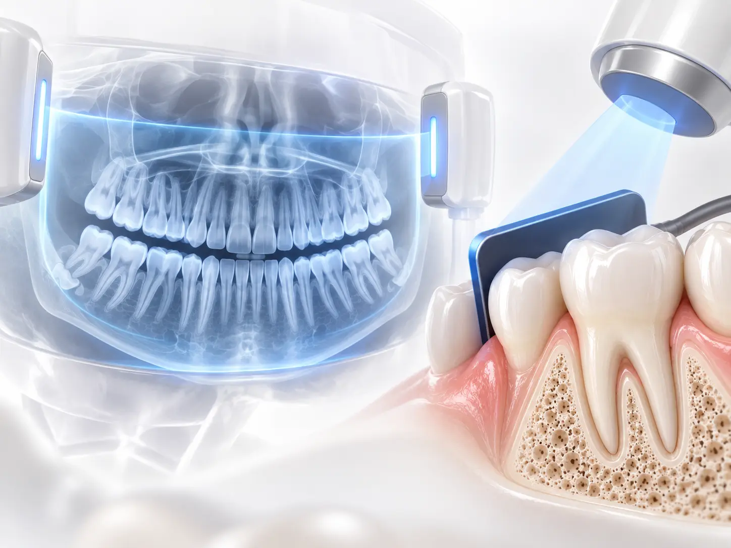
Panoramic and Periapical Imaging
A panoramic image covers a wider area, while a periapical image focuses on particular teeth and their roots. Other imaging may be considered when needed.
Interpreting the Images
Images are interpreted alongside clinical examination. The type and frequency of imaging are not the same for every patient.
Images are interpreted alongside the clinical examination; decisions are not based on an image alone.
What Happens at the First Consultation?
- 01
Your concerns, expectations and medical history are discussed.
- 02
A clinical examination is carried out and relevant diagnostic records are reviewed.
- 03
Treatment options, stages, possible risks and maintenance needs are discussed.
Suitability and a definitive plan are determined after clinical assessment. You can share existing X-rays and a list of medication before the visit.
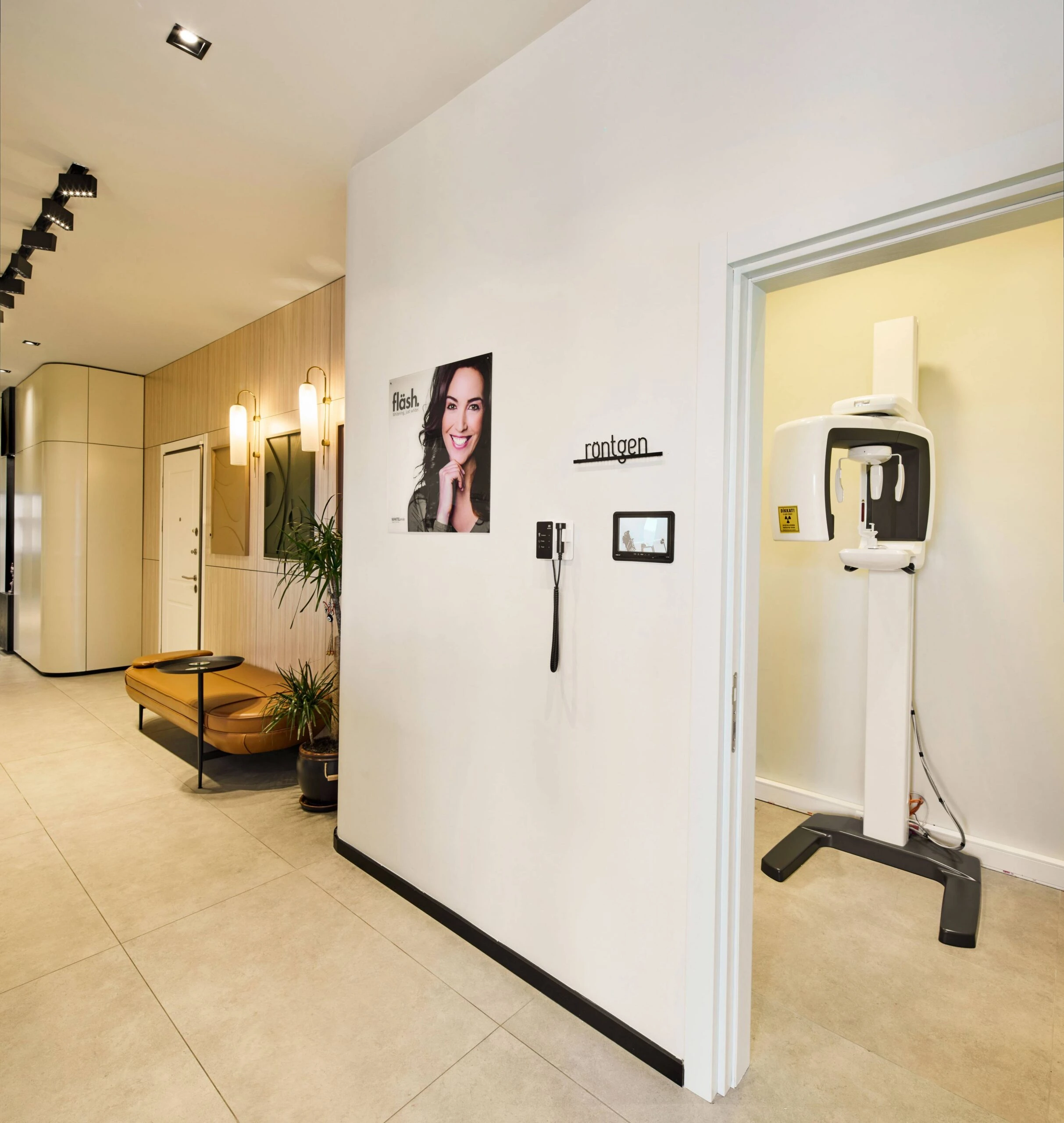
A Look Inside Our Practice



Panoramic and Periapical X-rays — Frequently Asked Questions
What information can these X-rays provide?
Panoramic and periapical X-rays provide different types of information during dental assessment. Imaging is selected according to examination findings and clinical need.
How is the type of image selected?
Previous images, symptoms and planned treatment are considered. Relevant health circumstances should be discussed before imaging.
How are images taken and interpreted?
A panoramic image covers a wider area, while a periapical image focuses on particular teeth and their roots. Other imaging may be considered when needed.
When would imaging be repeated?
A repeat depends on the adequacy of existing images and any new clinical question.
Is anaesthesia needed?
A standard dental examination or X-ray usually does not require anaesthesia; any other procedure is planned separately.
Can previous images be used?
Existing images are reviewed for date and diagnostic quality before a repeat is considered.
Can I have an examination without an X-ray?
Assessment begins with a clinical examination; imaging is chosen when needed for diagnosis or planning.
How are the images reviewed?
Images are interpreted alongside clinical examination. The type and frequency of imaging are not the same for every patient.
How do panoramic and periapical X-rays differ?
A panoramic view covers a broader area; a periapical image focuses on individual teeth and nearby tissues.
Can I send existing images from abroad?
Yes, they may inform a preliminary discussion; in-person assessment may still be needed.